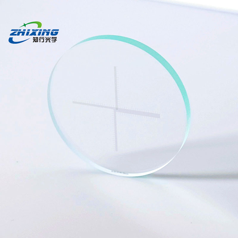
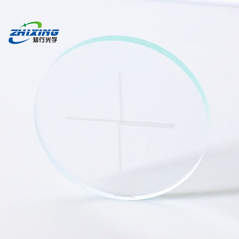
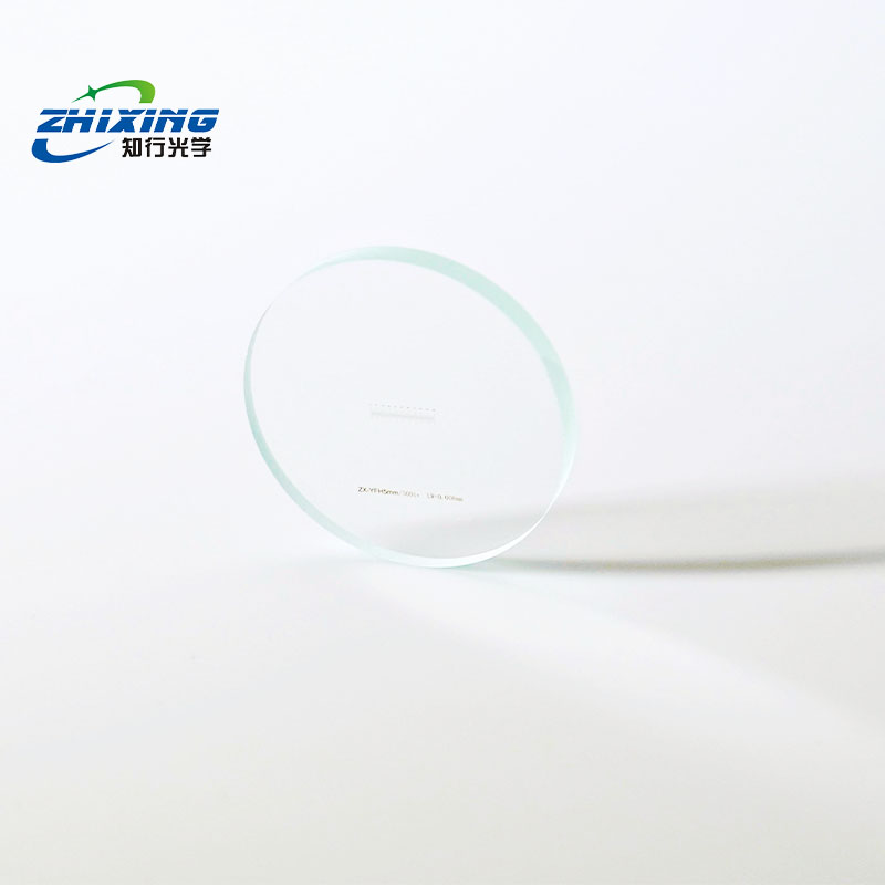
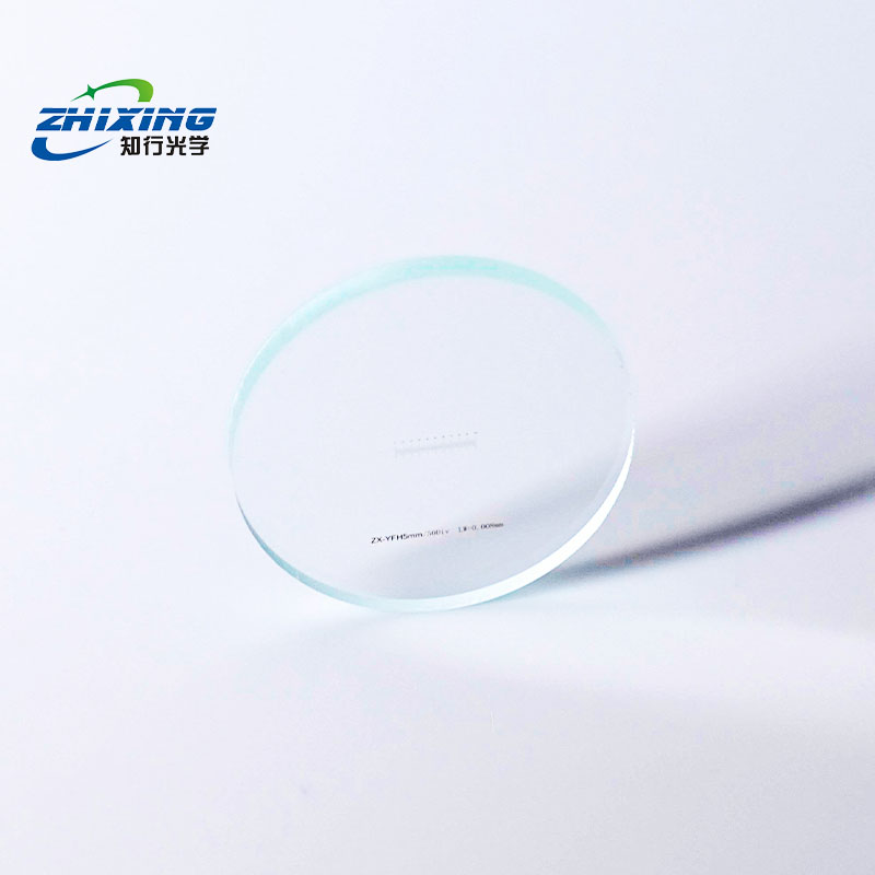
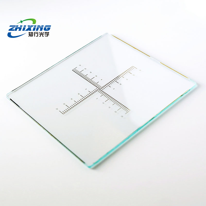
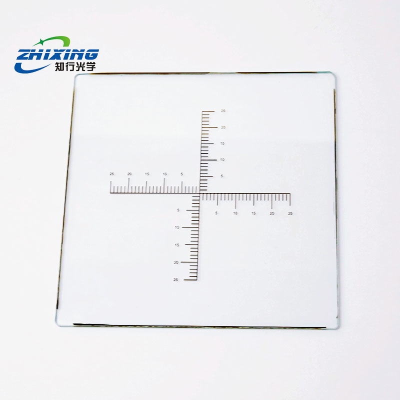
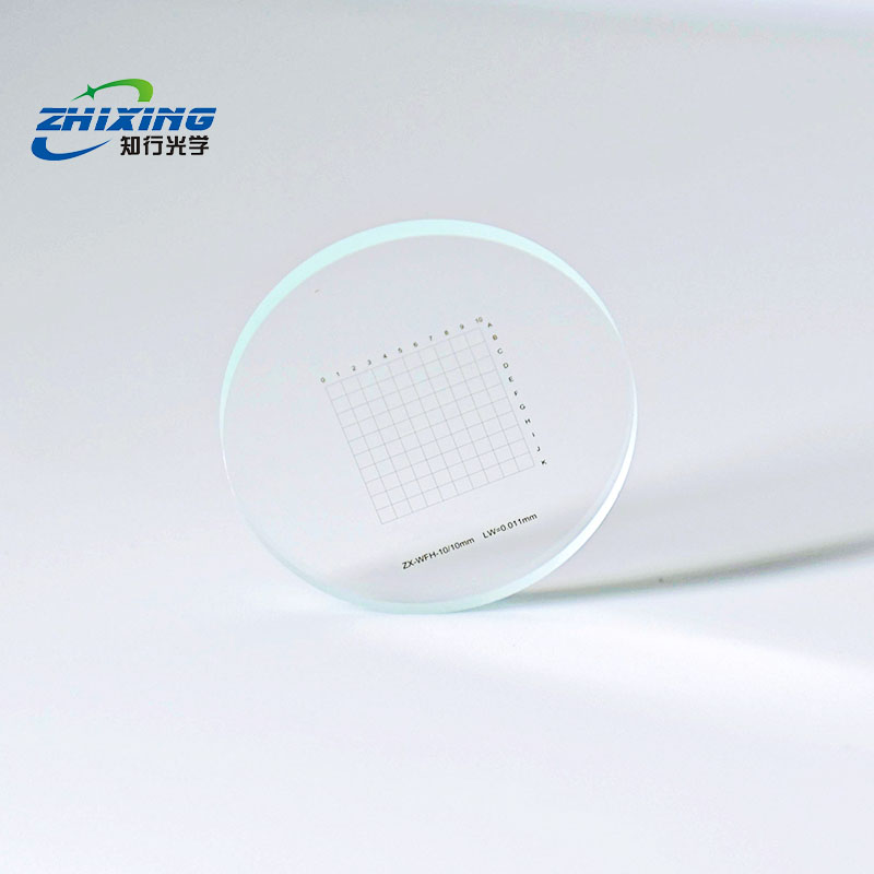
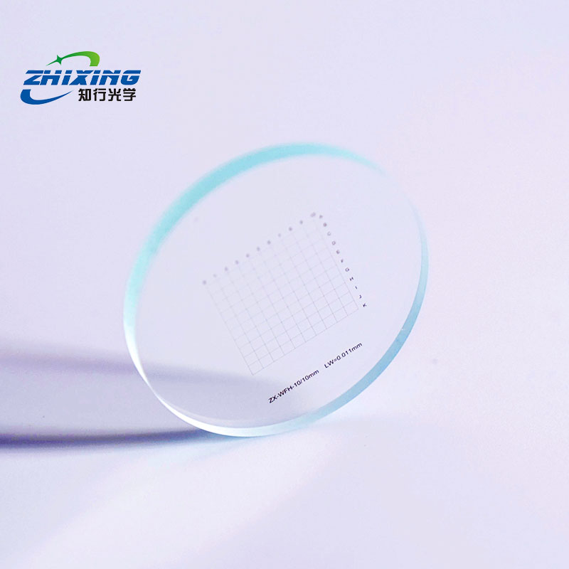
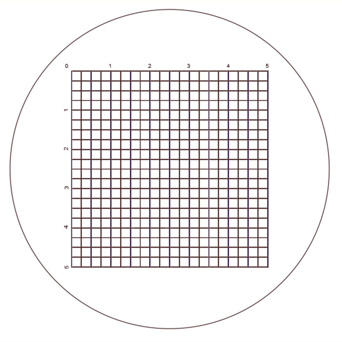
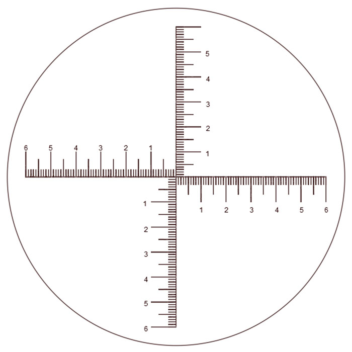
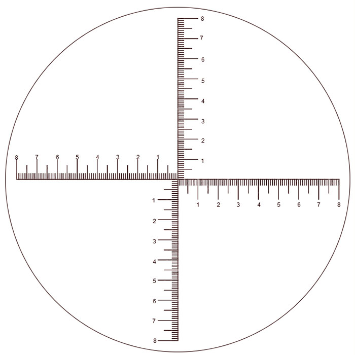
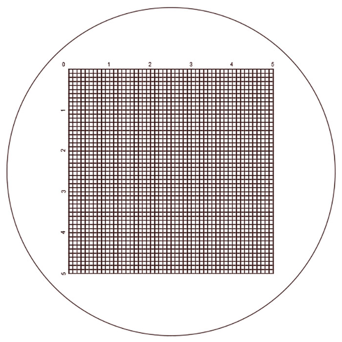
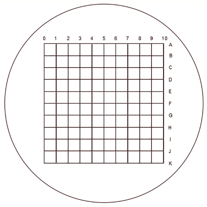
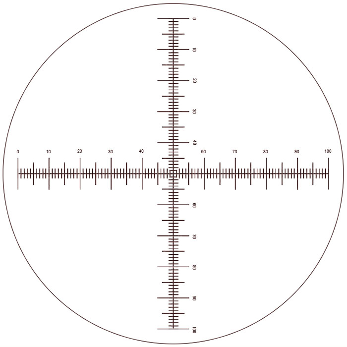
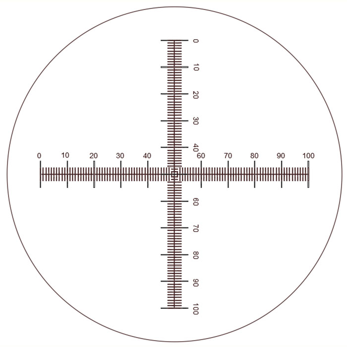
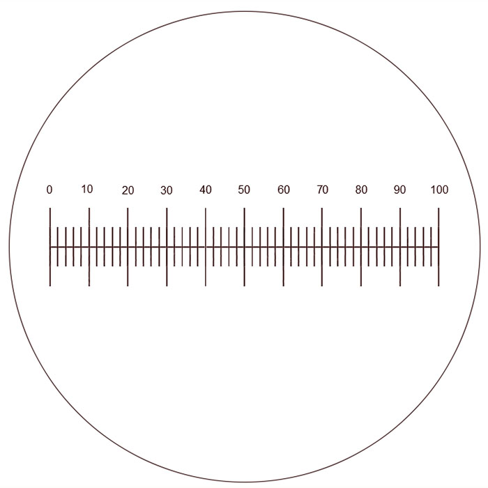
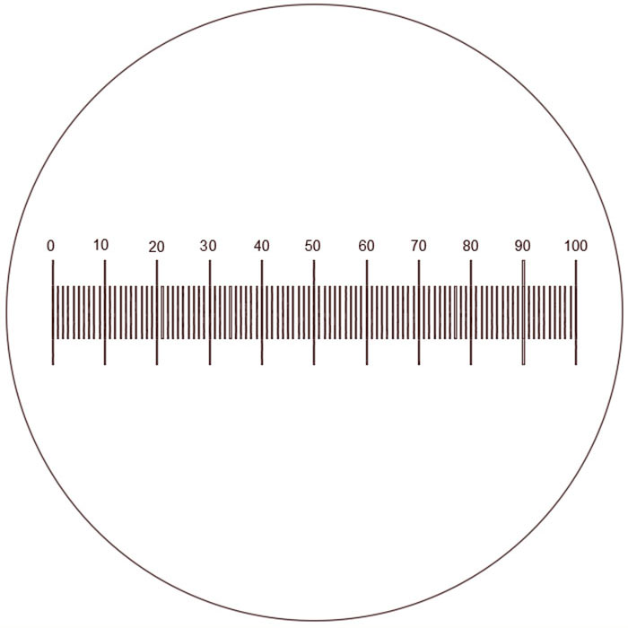
Eyepiece reticles (including linear scales, crosshair grids, cell counting chambers, etc.) are relative measurement tools when used under a microscope. Before use, they must first be "calibrated" using a stage micrometer at the current magnification to determine the calibration factor before accurate measurements can be made.
Step 1: Component Installation
Remove the Eyepiece: Take the microscope eyepiece out of the tube and unscrew the retaining ring (or clip) at the bottom of the eyepiece.
Placement: Wear finger cots or use professional tweezers to hold the reticle by its edges. Place it steadily into the focal plane (field stop) inside the eyepiece with the scale side facing down, then re-tighten the retaining ring.
Verify Field of View: Reinsert the eyepiece into the tube. Rotate the eyepiece or adjust the diopter until the black scale lines or grid lines appear sharp and clear in the field of view, ensuring that any numerals are not inverted.
Step 2: Magnification Calibration — Core Step
Note: This step must be repeated every time the objective magnification is changed.
Place a standard stage micrometer on the microscope stage and focus until the image is sharp.
Move the stage micrometer and rotate the eyepiece so that the reference starting point of the eyepiece reticle (e.g., the "0" mark or the top-left vertex of the grid) aligns perfectly with the "0" line of the stage micrometer.
Look outward to find the second point where the lines coincide exactly. Record the number of divisions on both scales within this interval.
Calculate the calibration factor per division:
Linear Scale: Calculate the actual length in micrometers represented by each small division on the eyepiece reticle.
Grid Scale: Calculate the actual physical dimension corresponding to "the side length of each square" or "the area of each grid unit."
Step 3: Specimen Observation
Remove the stage micrometer and replace it with the actual specimen slide to be examined. (Do not change the microscope magnification.)
Move the sample so that the target is positioned within the measurement area of the reticle:
Using a Linear Scale: Directly read how many small divisions the target object spans (used for measuring length, major/minor axes, or particle diameter).
Using a Grid Scale: Use the grid lines to perform regional counting (e.g., cell counting), multi-point statistics, or area coverage assessment on the target.
Step 4: Data Conversion
Actual Measurement = Reading from Eyepiece Reticle × Calibration Factor determined in Step 2
When recording data, always specify the objective magnification currently in use (e.g., 10×, 40×).
Product Selection Tips for Different Specifications
If you choose the [Standard Linear Scale]: Suitable for measuring the length, major/minor axes, or particle diameter of linear elastic objects. Quick readings can be obtained simply by aligning the line segments.
If you choose the [Grid/Mesh Type]: Specifically designed for cell counting, particle density analysis, and multi-level area distribution assessment. It allows statistical analysis of multiple target populations within a single field of view without the need for frequent sample translation.
Maintenance and Care Instructions
Strictly Avoid Touching with Bare Hands: Any fingerprints or sweat stains will compromise the clarity of the high-precision chrome-etched lines.
Cleaning Protocol: If dust is present on the surface, use a professional air blower bulb to remove it. For stubborn stains, gently wipe in one direction using an absorbent cotton ball moistened with a small amount of absolute ethanol. Never rub back and forth with excessive force.